SPECT - is a tomographic nuclear medicine imaging technique using gamma rays emitted from injected radionuclides. It is very similar to conventional nuclear medicine planar imaging using a gamma camera. However, it is able to generate a 3D tomographic data over the volume. The image data is typically displayed in transaxial, coronal, and sagittal views.
SPECT imaging, like PET, uses radioactively tagged tracer molecules to create functional images on the biochemistry or physiology of the subject. The specific biochemistry elucidated by the image depends upon the radiotracer. A SPECT radiotracer can image regional perfusion, specific receptor systems, organ function, or a number of other physiologically specific functions.
SPECT imaging can be quantitative in nature. Typically it is a quantitated relative uptake rather than absolute quantitation.
MicroSPECT small animal imaging can achieve resolutions of close to 1 mm.
 Micro Single Photon Emission Computed Tomography Equipment
 Micro Single Photon Emission Computed Tomography Equipment
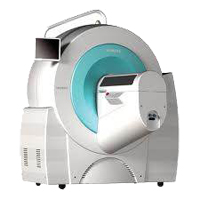
Siemens Inveon high resolution SPECT
- 2-head configuration with large active area 150 mm x 150 mm detector
- NaI crystals (2mm x 2mm x 10 mm) provide fine resolution
- Designed to detect gamma emissions from 30 keV-300 keV.
- Cardiac and Respiratory gating available
- SPECT and CT are within the same gantry
- This provides a co-registered anatomic image
Collimators available
- LEAP (low-energy all-purpose) parallel hole collimator for planar imaging
- Single pinhole includes either 0.5, 1.0, 2.0, or 3.0 mm sized pinhole
- 0.5 mm multi- pinhole for mouse brain
- 1.0 mm multi-pinhole for mouse whole body