Iowa Thoracic Surgery Protocols
see: Video of Thoracoscopic Ligation of the Thoracic Duct - Technique of 'Mass' Ligation
Please direct questions and comments to Evgeny V. Arshava, MD evgeny-arshava@uiowa.edu
See other sections in Iowa Thoracic Surgery
Esophageal Diverticula (Midesophageal and Epiphrenic): Esophageal Myotomy and Diverticulectomy
GENERAL CONSIDERATIONS
See: Chylothorax: diagnosis and management; Thoracic duct: anatomy and physiology
Indications
-
A chylous output of >10 mL/kg on the 5th day or >13 mL/kg/day on the 3rd day of nonoperative management (Dugue 1998, Miao 2015)
-
Increase in chyle output after reinstitution of oral diet or tube feedings.
Contraindications
*** Severe malnutrition is not a contraindication for the operation, as operation itself is intended to prevent malnutrition and lymphocytopenia.
-
Failed prior surgical TD ligation (relative)
-
Unstable spine injuries or severe multiple organ injuries (relative)
-
Hostile chest (multiple previous intrathoracic operations, prior radiation)
-
Severe comorbidities precluding single lung ventilation
-
Locally unresectable advanced tumor
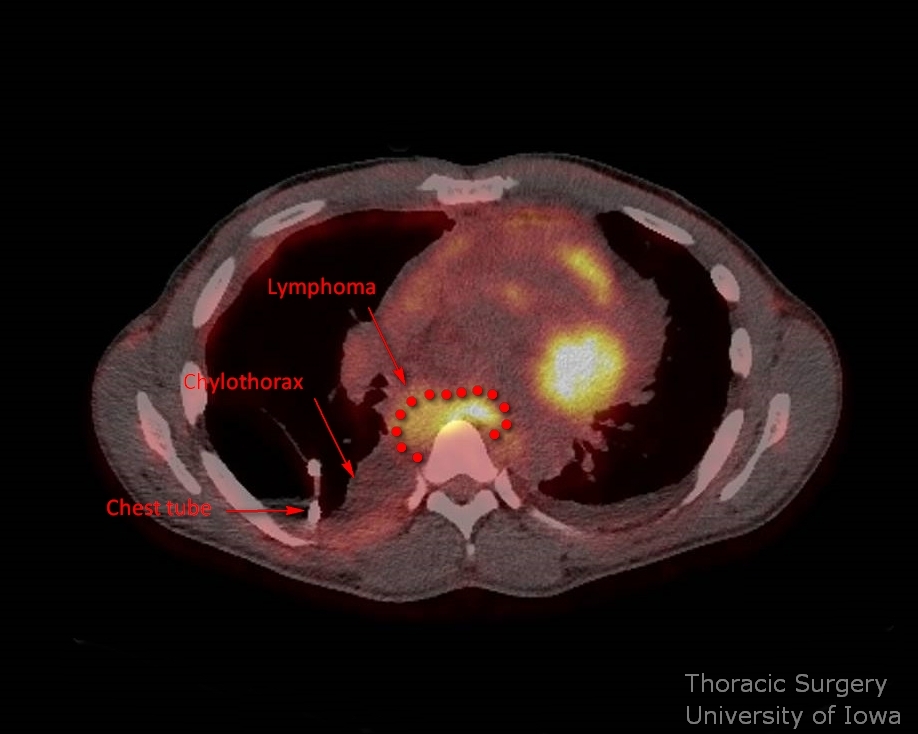
Percutaneous embolization
-
Surgery remains the cornerstone in the management of chylothorax refractory to medical management. TD ligation is effective in >90 % of cases with minimal morbidity of Video assisted thoracoscopic surgery (VATS).
-
In recent years percutaneous image guided TD embolization has been increasingly utilized and, in some centers, has reduced the use of surgical TDL. Percutaneous embolization is a time-consuming and difficult procedure even for an experienced interventional radiologist.
-
While reports indicate successful embolization in >70% of cases, this experience is limited to a few centers and should not be considered as a routine approach.
-
Diagnostic lymphangiography is easier to perform and may help to identify the location of the leak and select the surgical approach.
-
Lymphography and PE should be considered in following situations:
-
Suspected intraabdominal leak from CC and abdominal branches.
-
Failed TDL (relative)
Potential Complications
-
Failure to control chyle leak
-
Bleeding (azygous vein, aorta, intercostal vessels)
-
Injury to esophagus
-
Injury (or devascularization) of gastric conduit in cases of post-esophagectomy chylothorax
PREOPERATIVE PREPARATION
-
In most cases of postoperative chylothorax (esophagectomy and lung resection), the patient has already undergone a major operation and presumably appropriate preoperative cardiopulmonary work-up.
-
In cases of malignant chylothorax, appropriate cardiopulmonary evaluation for general anesthesia with single-lung ventilation is required.
-
As no lung needs to be resected even marginal pulmonary function tests ( PFT )are acceptable and are not needed.
ANESTHESIA CONSIDERATIONS
-
Double-lumen endotracheal tube
-
Paravertebral block versus intercostal block versus epidural (in cases of high likelihood of conversion to thoracotomy)
-
Two large-bore peripheral intravenous (IV) accesses
-
Radial arterial line optional
-
Blood type and cross
-
IV antibiotic within 1 h of the incision
-
Left decubitus position on a bean bag
NURSING CONSIDERATIONS
Instruments and Equipment
Standard
-
Major chest tray
-
Thoracoscopic equipment and tray
Special
-
Knot pusher
-
Endoscopic clip applier (optional)
Prep and Drape
-
Standard prep using chlorhexidine solution
-
Field includes supraclavicular area to below costal margin, spine to nipple line
-
Transparent incise drape (optional)
Drains and Dressings
- 28 or 32 Fr chest tube
OPERATIVE PROCEDURE
Operative approach
-
It is difficult and may be impractical to locate the exact site of the leak in the chest without lymphography. It is not necessary in most cases.
-
Regardless of laterality of the chylothorax, ligation of the thoracic duct (TD) at its proximal intrathoracic portion through the right chest will effectively controls chylothorax in most cases. It is essential to ligate the TD caudal to the leak and incorporate all potential collaterals.
-
Exploration and direct ligation of the leaking lymphatics could be considered after trauma through the left chest when non-urgent operation is needed for other reasons (retained hemathorax, lung hernia).
-
During esophagectomy areas where lymphatics are prone to injuries are cisterna chyle, large tributaries, and the TD as it crosses to the left side.
-
TDL for the chylothorax after a right transthoracic esophagectomy should be performed as the reoperation on the same side regardless of the leak laterality. Retraction of the conduit is not difficult with appropriate lung isolation.
-
Leaks with high suspicion of originating below the diaphragm may be best approached through the abdomen. Therefore, chylothorax after a difficult dissection around the hiatus are best localized preoperatively with lymphangiography.
-
Administering a high-fat substrate (olive oil or cream) by mouth or through NG or Dobhoff feeding tube if patient is strict NPO prior to operation may be helpful if an attempt of direct repair is planned (like penetrating trauma or postoperative neck reexploration).
-
Injection of methylene blue or other dye is not needed. This will stain the entire chest and make dissection more difficult.
-
For cases of spontaneous chylothorax, pleural biopsies should be performed to rule out malignant processes.
Thoracoscopic thoracic duct ligation
-
Thoracoscopic ports are typically placed in the following manner:
-
Camera site: 7th or 8th intercostal space on the posterior axillary line.
-
Working incision #1: 5th or 6th intercostal space on posterior axillary line.
-
Working incision #2: 8th or 9th space posterior to the camera incision.
-
Optional working port #3 may be placed, but rarely needed
-
-
Both the surgeon and assistant stand on the left side of the table with assistant holding the 30o camera.
-
The diaphragm may be pushed down with a sponge stick or ring forceps through the camera port.
-
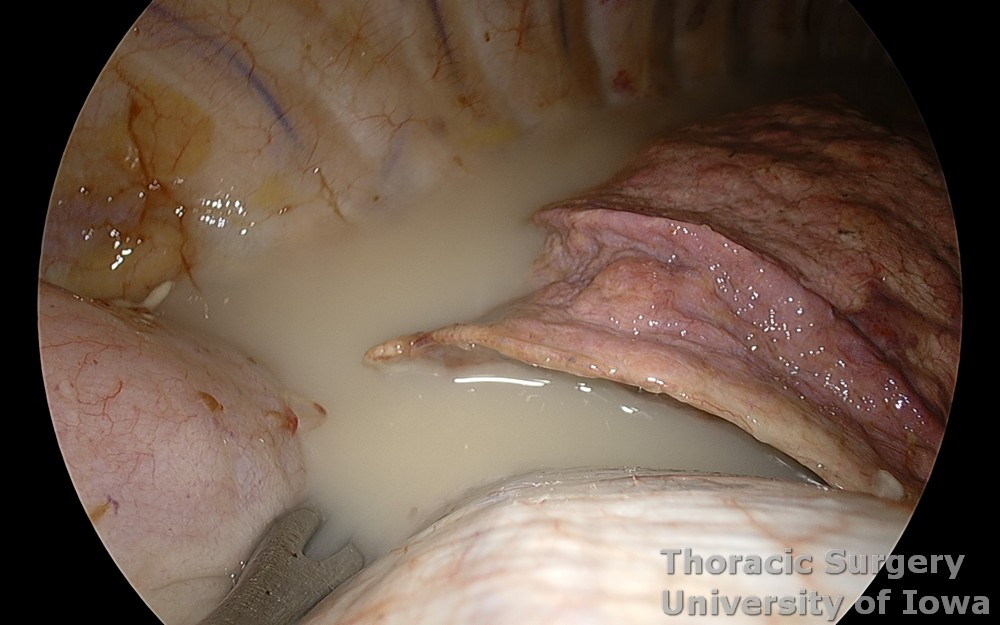
The chest is inspected and undrained chylothorax evacuated
-
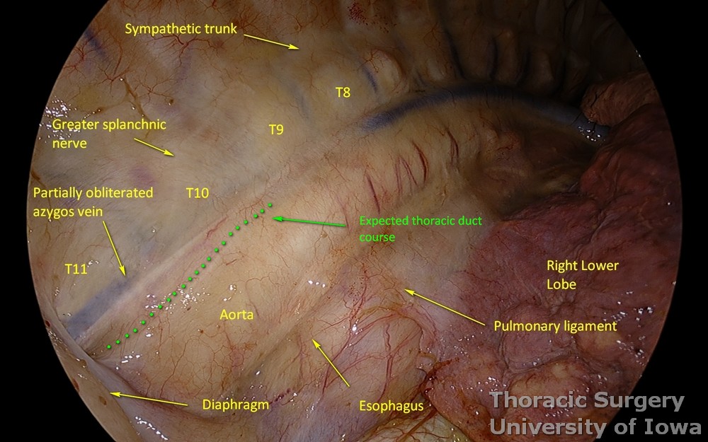
The deflated lung is retracted anteriorly and cephalad for exposure of the posterior mediastinum.
-
The pulmonary ligament division to improve exposure is rarely needed
-
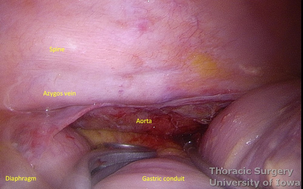
If TD ligation is performed for postoperative chylothorax after the esophagectomy, the gastric conduit is identified.
-
If an additional exposure needed, the gastric conduit must be retracted with care to the left of the spine to avoid trauma and undue traction onto the gastroepiploic pedicle.
-
The site for TD ligation is selected typically at the level of the inferior ligament (T10-11). Most abdominal tributaries are expected to already join TD below this level.
-
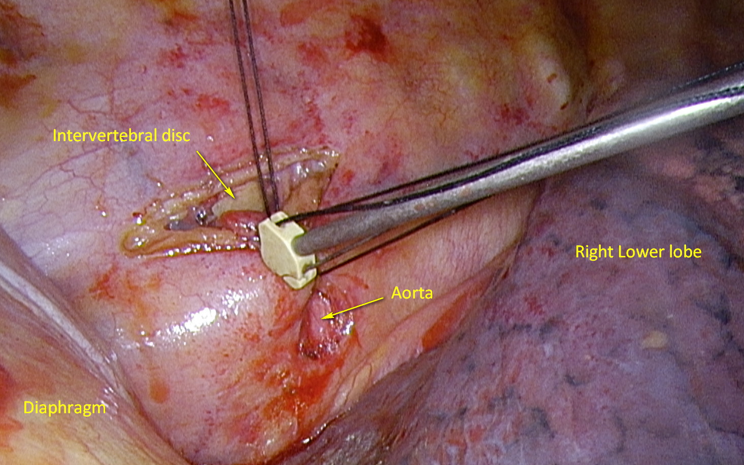
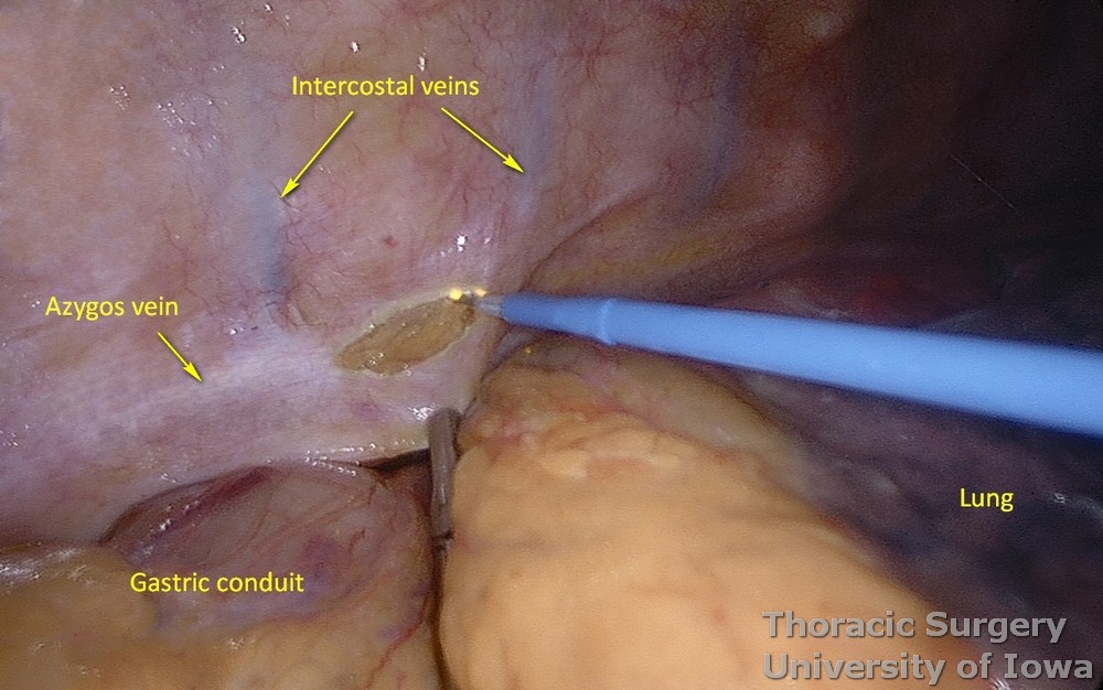
The parietal pleura is incised lateral to the azygous vein to clear a 2 cm area over a prominent intervertebral disk.
-
Intercostal veins are located over the vertebral bodies and usually do not need to be divided.
-
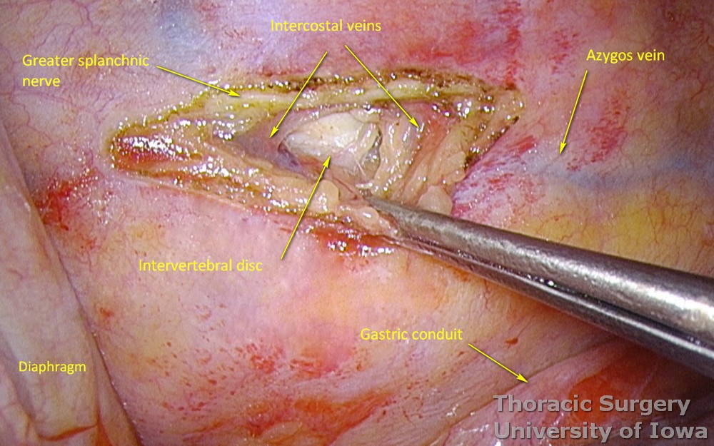
The intervertebral disk is visualized. The greater splanchnic nerve is preserved, while segmental branches may be divided medial to it.
-
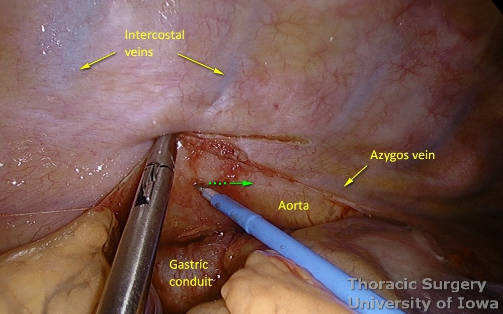
The aorta is identified visually or by gentle palpation with the dissecting instrument in obese patients or if tissues are edematous. The parietal pleura is incised now over the projection of right lateral border of the aorta.
-
Using right angle clamp or Harken dissector, all the tissue above the aorta are dissected off the spine.
-
First gently dissect with the tip of the instrument along aorta and then slide it perpendicular onto the spine.
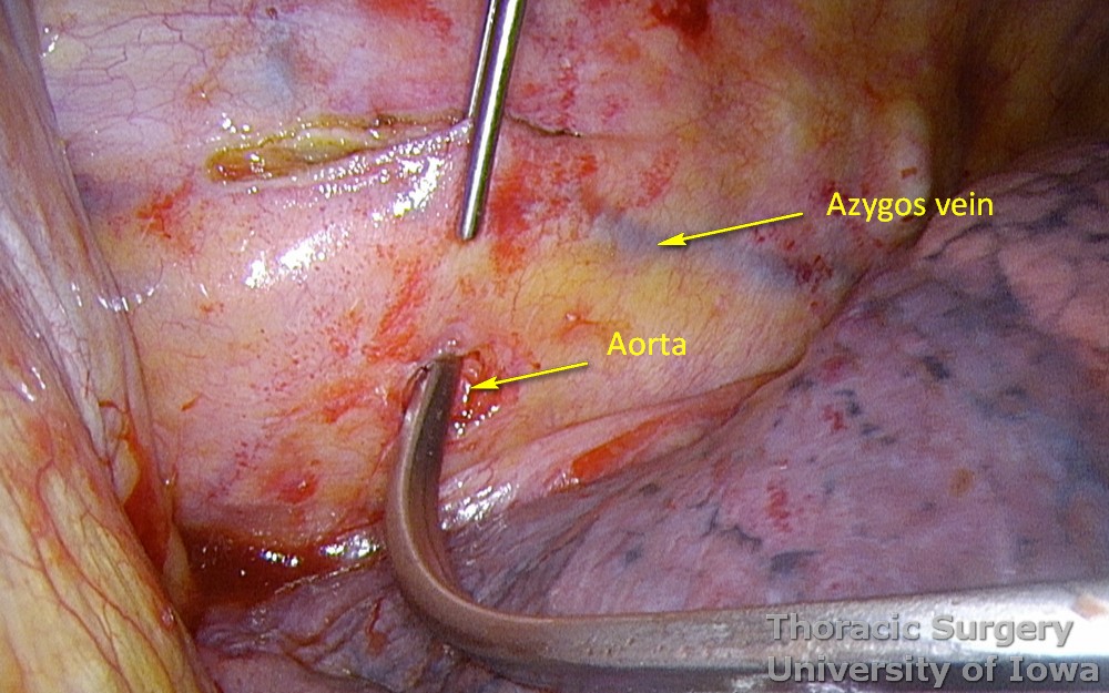
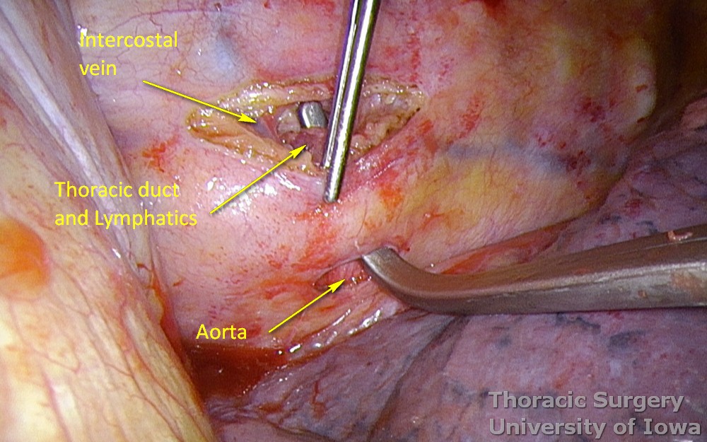
-
These tissues (including the azygos vein, TD, potential lymphatic branches, parietal pleura) are encircled with a silk tie.
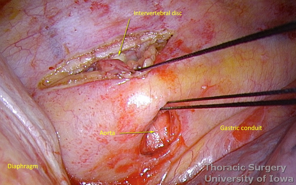
-
Tactile feedback while gently palpating aorta and spine with the tip of the dissecting instrument is very helpful to assure all the lymphatics are dissected between the spine and aorta.
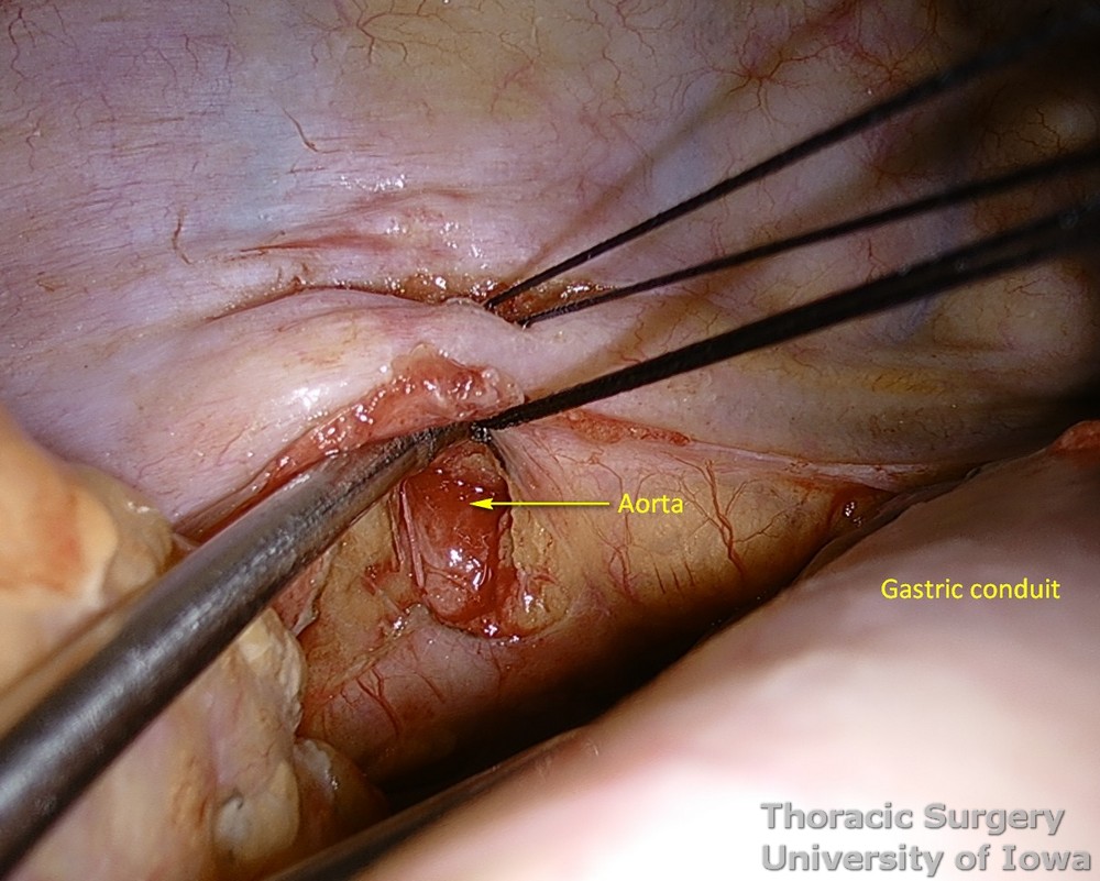
-
Protect intercostal arteries.
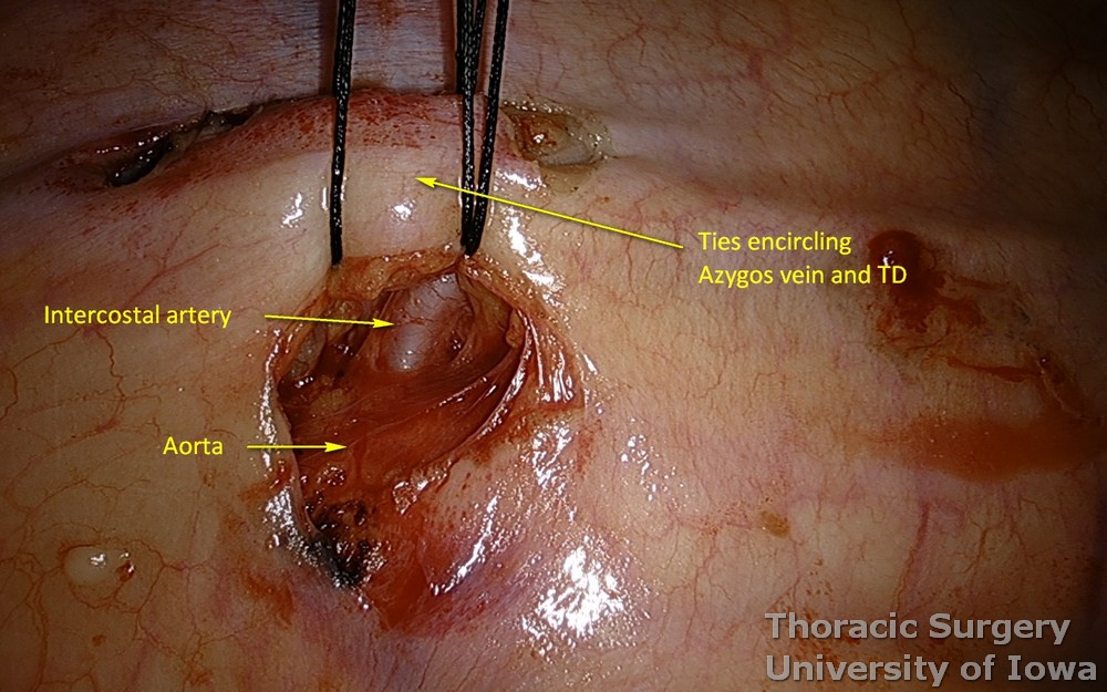
-
Usually two or three passes of the dissector are needed to dissect and encircle all lymphatics (potential branches). Gentle traction on silk facilitates subsequent passes.
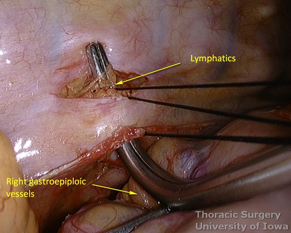
-
Silk sutures are tied using a knot pusher.
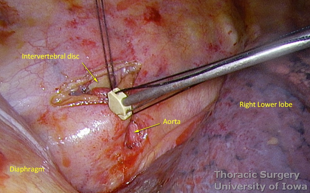
-
After tying the ligatures, NO division of the duct is performed.
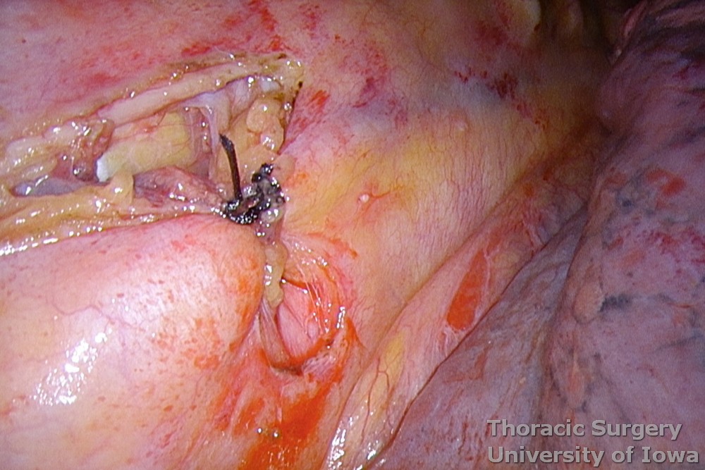
-
Clips may be very selectively added as well but are not needed in most cases if the dissection was not traumatic.
-
Existing metal clips most commonly, however, do not necessarily encompass the thickness of the dissected tissue, especially in obese patients. Larger plastic hemoclips close too tight and may damage the duct.
-
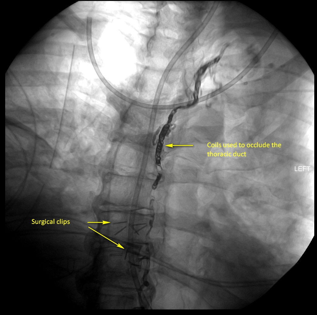
One must not rely on the number of clips used. They will fail unless all tissues between aorta, spine, and azygos vein are ligated. Below is the image of the percutaneous embolization of the thoracic duct after failed surgical ligation and talc pleurodesis in an outside institution. Note multiple surgical clips that failed to control the thoracic duct.
-
We feel that the technique to dissect the TD individually may lead to duct injury in frequently inflamed planes and to miss a potential duplication or large branch. Including the azygos vein and parietal pleura to the mass ligation adds an additional buttressing strength to the tie.
-
A chest tube is placed through the camera port site.
Thoracic duct ligation via thoracotomy
-
Thoracotomy may be necessary if thoracoscopic expertise is not available or in cases when thoracoscopy is not safe due to inflammatory changes or prior operations.
-
In all other cases VATS offers much better visualization of the area.
-
A right posterolateral thoracotomy is performed through the 6th or 7th intercostal space with division of latissimus dorsi muscle and preservation of serratus anterior muscle.
-
A heavy stitch maybe placed onto the dome of the diaphragm to retract it through a future chest tube site.
-
Digital palpation is very helpful to identify the aorta and spine, and to assure that all tissues between them are included in ligation.
-
Other steps are identical to VATS TD ligation.
-
A chest tube is placed and ribs are approximated with figure of eight #2 absorbable pericostal stitches on a blunt needle. The thoracotomy is closed in layers with absorbable suture.
-
Skin is closed with a running subcuticular stitch. Surgical dressing or skin adhesive is used to cover incision.
POSTOPERATIVE CARE
General
-
Patient is extubated in the operating room and admitted to a regular surgical ward.
-
Aggressive pulmonary care:
-
Ambulation at least four times daily is recommended.
-
Incentive spirometer every our hour.
-
Vibrating Positive Expiratory Pressure therapy every our hour.
-
Scheduled bronchodilators every 4 hours
-
Scheduled chest physical therapy with the use of manual Pneumatic Percussor on both lung fields every 4 hours while awake. Manual percussion even of the operative side is well tolerated by patients and may be more effective than a built in percussion function of hospital beds.
-
Use mucolytics inhalers as needed
-
-
Patient after esophagectomy are prone to aspiration (lack of gastric storage capacity, removal of LES and nonfunctional pylorus). Head of the bed elevation, small meal are standard precautions.In cases of the postoperative ileus a functioning NG is aimed to fully decompress gastric conduit.
-
Postoperative prophylactic antibiotics are not indicated.
-
Prophylactic heparin may be administered on POD 0.
Specifics
-
Oral / enteral intake is resumed the following day.
-
A chest x-ray (CXR) is obtained to assure full re-expansion of the lung on postoperative day 1.
-
Chest tube is maintained on suction at –20 cm H2O (off suction for ambulation) for first 1-2 days and then water sealed. No ongoing chyle leak is expected after TD ligation. The chest tube can be removed when output is < 200 mL of serous fluid per day while the patient is receiving a regular diet or tube feedings.
OUTPATIENT FOLLOW-UP
-
A CXR should be performed 1–2 weeks after chest tube removal to assure lack of effusion reaccumulation and full expansion of the lung.
REFERENCES
Cushing HW. Operative wounds of the thoracic duct: report of the case with suture of the duct. Ann. Surg. 27:719, 1898.
Zesas DG. Die nicht operativ entstandenen Verletzungen des Ductus thoracicus. Deutsch. Z. Chir. 115:49, 1912 [in German].
Иосифовъ ГМ. Лимфатическая система человѣка съ описанiемъ аденоидовъ и органовъ движенiя лимфы. Съ 80 рис. въ текстѣ и таблицахъ. Извѣстiя Императорскаго Томскаго Университета. 1914; 59:3:1-96 [in Russian]
Davis H. A statistical study of the thoracic duct in man. American Journal of Anatomy. Jan. 1915
Zhdanov DA. The functional anatomy of the lymphatic system. Gorky, The Soviet Union: Gorky State Medical Institute; 1940. [in Russian]
Lampson RS. Traumatic chylothorax; a review of the literature and report of a case treated by mediastinal ligation of the thoracic duct. J Thorac Surg. 1948 Dec;17(6):778-91
Klepser RG, Berry JF. The diagnosis and surgical management of chylothorax with the aid of lipophilic dyes. Diseases of the chest 25:409, 1954.
Defize IL, Schurink B, Weijs TJ, Roeling TAP, Ruurda JP, van Hillegersberg R, Bleys RLAW. The anatomy of the thoracic duct at the level of the diaphragm: A cadaver study. Ann Anat. 2018 May; 217:47-53. doi: 10.1016/j.aanat.2018.02.003. Epub 2018 Mar 3.
Johnson OW, Chick JF, Chauhan NR, Fairchild AH, Fan CM, Stecker MS, Killoran TP, Suzuki-Han A. The thoracic duct: clinical importance, anatomic variation, imaging, and embolization. Eur Radiol. 2016 Aug;26(8):2482-93. doi: 10.1007/s00330-015-4112-6.
Kent RB 3rd, Pinson TW. Thoracoscopic ligation of the thoracic duct. Surg Endosc. 1993 Jan-Feb;7(1):52-3.
Hou X, Fu JH, Wang X, Zhang LJ, Liu QW, Luo KJ, Lin P, Yang HX. Prophylactic thoracic duct ligation has unfavorable impact on overall survival in patients with resectable oesophageal cancer. Eur J Surg Oncol. 2014 Dec;40(12):1756-62.
Miao L, Zhang Y, Hu H, Ma L, Shun Y, Xiang J, Chen H. Incidence and management of chylothorax after esophagectomy. Thorac Cancer. 2015 May;6(3):354-8.
