Holden Comprehensive Cancer Center is among the first to adopt technology blending high-quality MR imaging with radiation therapy.
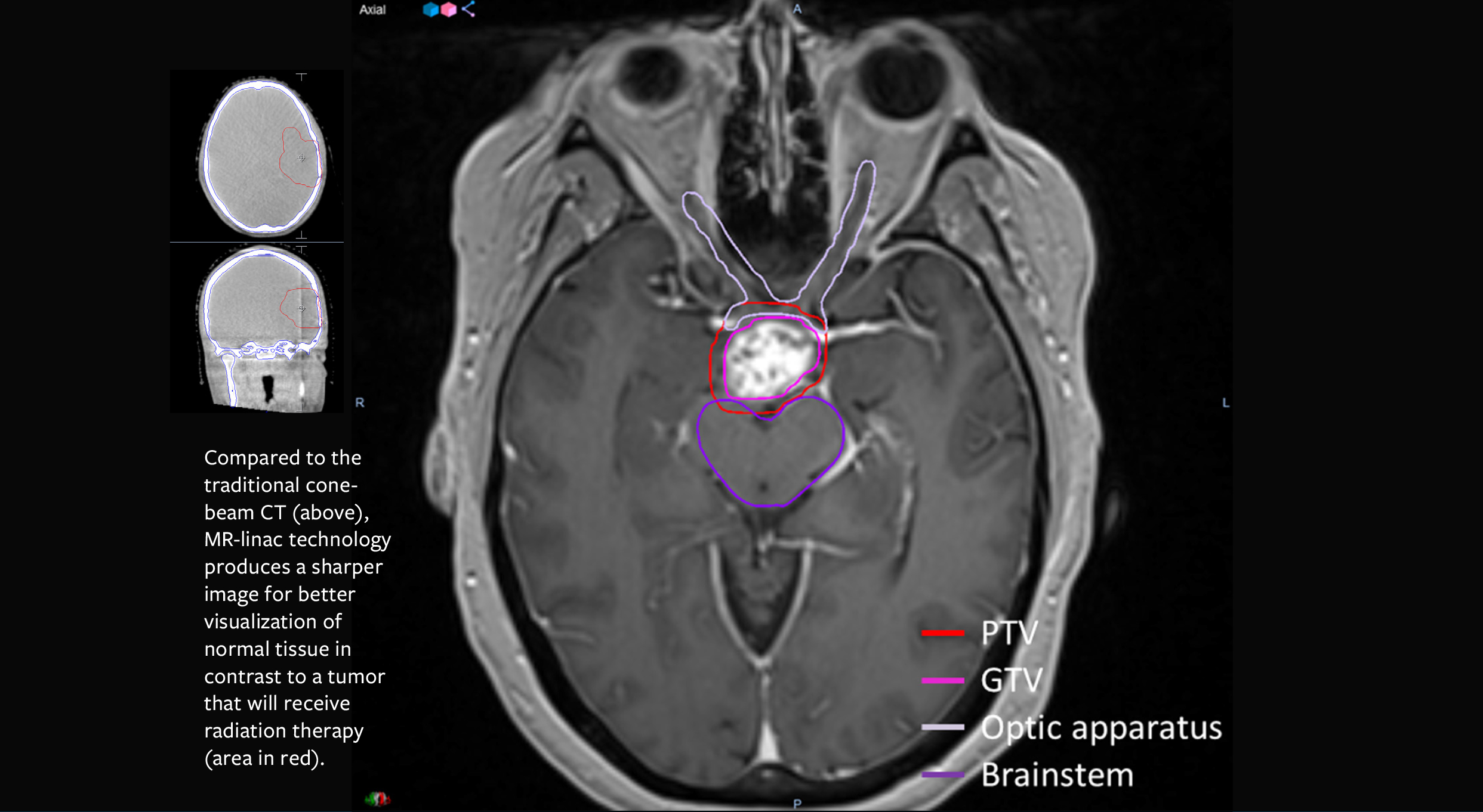 A new radiation therapy device, MR-linac, combines state-of-the-art magnetic resonance (MR) imaging with radiation treatment to enable unparalleled accuracy in attacking tumors. For providers, the difference between a scan using traditional cone-beam CT and the MR-linac is like a farsighted person putting on glasses to improve image sharpness, definition, and clarity of detail.
The National Cancer Institute-designated Holden Comprehensive Cancer Center at the University of Iowa is one of the first three U.S. cancer centers to start treating cancers with MR-linac technology containing a 1.5 Tesla MRI scanner. The arrival of MR-linac at Iowa has the potential to greatly improve outcomes for patients with cancers including tumors on the prostate, brain, pancreas, liver, breast, and lung.
“The ability to view tissue with MR-linac versus CT is remarkable. The high-Tesla magnet combined with linac provides significantly better images than previous technology,” says John Buatti, MD, chair of the Department of Radiation Oncology and a Holden Comprehensive Cancer Center member.
Buatti leads the team using MR-linac technology to personalize treatment of cancer. The machine, which started treating patients in May, will eventually treat approximately 175 patients each year as the therapy becomes more widely known.
Success hitting moving targets
Within the next year, the MR-linac will also allow doctors to gate the radiation therapy with unprecedented accuracy, which is especially important for tumors than can easily change position. The MRI-linac imaging produces five images per second, with significantly better soft-tissue contrast than the cone-beam CT imaging used in traditional linear accelerator radiation therapy, so visualization of tumors and surrounding normal tissue is vastly improved.
Care teams use MR-linac to develop and modify treatment plans on a daily basis to treat tumors more precisely, without the extra dose of radiation that a cone-beam CT delivers. These personalized treatment plans are customized according to a tumor’s position, shape, biology, and relative location to sensitive organs. Each treatment is designed specifically for that patient that day.
The breakthrough in MR-linac technology is in its design, which overcomes the MRI magnet’s interference in the linear accelerator’s beam generation and, conversely, prevents the beam’s degradation of the MR image. A radio frequency “cage” prevents the two technologies from disrupting each other. By delivering higher doses of radiation more accurately, the therapy can kill tumor cells more efficiently with less exposure to surrounding tissue. This can mean fewer treatments for some patients.
“For example, we are treating a patient with a recurrent tumor at the base of the spinal cord. This technology improves our confidence level because we can shape the dose around the cord,” Buatti says.
Nancy Habinck, 73, had treatment for a rare chordoma nine years years ago. She had surgery to remove the tumor on her spinal column, as well as radiation, but it returned. This type of tumor is not treated with chemotherapy. Withthe MR-linac, Habinck had the benefit of improved treatment by imaging the dose of radiation in relationship with her spinal cord daily.
“I thought I couldn’t have any more radiation, but Dr. Buatti said the [MR-linac] technology was an option for me. If I couldn’t have this treatment, I might not be here,” Habinck says.
Habinck’s care with MR-linac involved twice-daily sessions, six hours apart, for a total of 50 treatments. Rather than make a daily drive from their home in Waterloo, Iowa, she and her husband stayed at Hope Lodge in Iowa City, near UI Hospitals & Clinics, for the duration of her treatment.
“Not many places can take care of this type of rare tumor,” Habinck says. “I’m so grateful to have this treatment available here in eastern Iowa.”
Holden team aims to sharpen accuracy even more
Buatti and a team of UI radiation oncologists and physicists were part of an international consortium of academic medical centers that collected data to prepare the MR-linac technology for effective clinical performance. Preparation and training to bring the treatment to Holden Comprehensive Cancer Center lasted more than a year.
As an academic medical center, UI radiation oncologists and physicists are not only using the new technology to treat patients; they are working to improve it. Buatti’s team has submitted a grant application to explore how verification of the dose using MR-linac is calculated. Currently, the machine is designed to recalculate the treatment dose daily, but the doses required can change with changes in position of the tissue. Buatti’s team has been developing a mathematical algorithm that will verify the dose much more quickly and improve dosing accuracy.
Buatti’s team is also working toward obtaining regulatory approval to use the MR-linac technology to assess the functional nature of tumors and explore how they respond to treatment on a cellular level. The UI expects to become a training center for MR-linac technology and will teach others how to use the treatment technique as well.
By Amy Sundermann